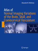
Atlas of Normal Imaging Variations of the Brain Skull and Craniocervical Vasculature 1st Edition by Alexander M Mckinney ISBN 3319397893 978-3319397894
$50.00 Original price was: $50.00.$25.00Current price is: $25.00.
Atlas of Normal Imaging Variations of the Brain Skull and Craniocervical Vasculature 1st Edition by Alexander M Mckinney – Ebook PDF Instant Download/Delivery:3319397893 ,978-3319397894
Full download Atlas of Normal Imaging Variations of the Brain Skull and Craniocervical Vasculature 1st Edition after payment
Product details:
ISBN 10: 3319397893
ISBN 13: 978-3319397894
Author: Alexander M Mckinney
This atlas presents normal imaging variations of the brain, skull, and craniocervical vasculature. Magnetic resonance (MR) imaging and computed tomography (CT) have advanced dramatically in the past 10 years, particularly in regard to new techniques and 3D imaging. One of the major problems experienced by radiologists and clinicians is the interpretation of normal variants as compared with the abnormalities that the variants mimic. Through an extensive collection of images, this book offers a spectrum of appearances for each variant with accompanying 3D imaging for confirmation; explores common artifacts on MR and CT that simulate disease; discusses each variant in terms of the relevant anatomy; and presents comparison cases for the purpose of distinguishing normal findings from abnormalities. It includes both common variants as well as newly identified variants that are visualized by recently developed techniques such as diffusion-weighted imaging and multidetector/multislice CT. Thebook also highlights normal imaging variants in pediatric cases.
Atlas of Normal Imaging Variations of the Brain, Skull, and Craniocervical Vasculature is a valuable resource for neuroradiologists, neurologists, neurosurgeons, and radiologists in interpreting the most common and identifiable variants and using the best methods to classify them expediently.
Table of contents:
Part I: Brain
1: Brain: Introduction
1.1 Terminology
2: Cerebellar Tonsillar Ectopia
Suggested Reading
3: Cerebellar Flocculus Pseudomass
Suggested Reading
4: Mega Cisterna Magna and Retrocerebellar Arachnoid Cysts
4.1 Retrocerebellar Arachnoid Cyst
4.2 Fetal MRI of the Normal Posterior Fossa Versus True Abnormalities
Suggested Reading
5: Cranial Nerve VII: Normal Contrast Enhancement on Magnetic Resonance Imaging
5.1 Vestibular Aqueduct Enhancement
Suggested Reading
6: Meckel Cave Prominence, Asymmetry, and Petrous Apex Cephaloceles
Suggested Reading
7: Cavernous Sinus Fat and Pseudomasses
7.1 Cavernous Sinus Pseudomass on CT
Suggested Reading
8: Pituitary Variations, Artifacts, Primary Empty Sella, and Incidentalomas
8.1 Lateral Anterior Lobe Pituitary Enhancement
8.2 Pituitary Hypertrophy and Tenting
8.3 Infundibular Shift or Tilt and Pituitary Asymmetry
8.4 Infundibular Hyperintensity on Three-Dimensional FLAIR
8.5 Artifacts Simulating Pituitary Microadenomas
8.6 Primary Empty Sella (PES): Partial Form
8.7 Rathke Cleft and Pars Intermedia Cysts
Suggested Reading
9: Liliequist Membrane
Suggested Reading
10: Pineal Gland: Normal Size, Appearance, and Enhancement
10.1 The Pineal Gland: Normal Calcifications
10.2 Pineal Cysts
Suggested Reading
11: Choroid Plexus: Normal Locations and Appearances
11.1 Choroid Plexus Cysts
11.2 Choroidal Fissure and Neuroepithelial Cysts
11.3 Choroid Plexus Xanthogranulomas
11.4 Choroid Plexus and Pericallosal Lipomas
11.5 Choroidal Fissure Asymmetry
Suggested Reading
12: Hippocampi, Caudate Tail, and Ependymal Variants
12.1 Hippocampal Cysts
12.2 Caudate Tail Pseudolesion and Hippocampal Anatomy
12.3 Hippocampal Corrugated Appearance
12.4 Periventricular Ependymal Pseudolesion, Ependymitis Granularis, and Connatal Cysts
References
Suggested Reading
13: Midline Variants of the Septum Pellucidum, Corpus Callosum, and Massa Intermedia
13.1 Cavum Septum Pellucidum, Cavum Et Vergae, and Cavum Velum Interpositum
13.2 Subcallosal/Paraterminal Pseudomass on Midline Sagittal Images
13.3 Massa Intermedia Pseudomass on Sagittal T1WI
13.4 Callosal-Septal and Pericallosal Pseudolesions on Sagittal FLAIR MRI
13.5 Callosal Isthmus
13.6 Callosal Vascular Indentation by the Anterior Cerebral Artery or Its Branches
Suggested Reading
14: Dilated Perivascular Spaces
14.1 Dilated Perivascular Spaces: Type I
14.2 Extensive Type 1 Dilated Perivascular Spaces: État Criblé
14.3 Dilated Perivascular Spaces: Type II
14.4 Dilated Perivascular Spaces: Type III
14.5 Tumefactive Perivascular Spaces
References
Suggested Reading
15: Enlargement or Asymmetry of the Lateral Ventricles Simulating Hydrocephalus
Suggested Reading
16: Cerebral or Cerebellar Volume Loss Simulating Subdural Hematomas, Hygromas, and Arachnoid Cysts
16.1 Cerebellar (Without Cerebral) Volume Loss Simulating Chronic Subdural Hematomas or Hygromas
16.2 Cerebellar and Cerebral Atrophy Simulating Chronic Subdural Hematomas or Hygromas
16.3 Cerebral (Without Cerebellar) Atrophy Simulating Chronic Subdural Hematomas or Hygromas
16.4 Enlarged Subarachnoid Spaces Simulating Cyst, Subdural, or Encephalomalacia
Suggested Reading
17: Dural Calcifications: Normal Locations and Appearances
Suggested Reading
18: Pseudo-leptomeningeal Contrast Enhancement
Suggested Reading
19: Basal Ganglia: Physiologic Calcifications
19.1 Basal Ganglia Calcifications: Unilateral or Asymmetric
Suggested Reading
20: Susceptibility-Weighted Imaging: Concepts, Basal Ganglia Variation in Age-Related Iron Depositio
20.1 Susceptibility-Weighted Imaging: Phase Maps
20.2 Susceptibility-Weighted Images: Normal Basal Ganglial Iron Deposition
20.3 Susceptibility-Weighted Images: Normal Iron Deposition at 0 to 10 Years of Age
20.4 Susceptibility-Weighted Images: Normal Iron Deposition at 10 to 20 Years of Age
20.5 Susceptibility-Weighted Images: Normal Iron Deposition at 20 to 40 Years of Age
20.6 Susceptibility-Weighted Images: Normal Iron Deposition at 40 to 60 Years of Age
20.7 Susceptibility-Weighted Images: Normal Iron Deposition at 60+ Years of Age
20.8 Susceptibility-Weighted Images: Artifacts Simulating Hemorrhages
20.9 Susceptibility-Weighted Images: Artifact from Air-Bone Interfaces and Gas
20.10 Susceptibility-Weighted Images: Artifacts from Metal Extensively Obscuring the Cranium
20.11 Susceptibility-Weighted Images: Motion-Related Artifact
20.12 Susceptibility-Weighted Images: Vasculature Simulating Microhemorrhages
20.13 Susceptibility-Weighted Images: Choroid Plexus Simulating Microhemorrhages
20.14 Susceptibility-Weighted Images: Postcontrast Appearance and the Use of Subtraction Imaging
Suggested Reading
21: Slow-Flow, Asymptomatic Vascular Malformations: Brain Capillary Telangiectasias and Development
21.1 Brain Capillary Telangiectasias
21.1.1 Brain Capillary Telangiectasias: Pontine
21.2 Capillary Telangiectasias: Cerebellar and Medullary
21.2.1 Capillary Telangiectasias: Basal Ganglial, Capsular, Frontal Basal, Insular, and Thalamic
21.2.2 Capillary Telangiectasias: Callosal and Cingulate
21.2.3 Capillary Telangiectasias: Lobar
21.3 Developmental Venous Anomalies
21.3.1 Developmental Venous Anomalies: Cerebellar
21.3.2 Developmental Venous Anomalies: Lobar
21.3.3 Developmental Venous Anomalies: Giant and Holohemispheric Types
21.3.4 Overlap Between Developmental Venous Anomaly and Capillary Telangiectasia
21.3.5 Comparison Cases of Symptomatic Developmental Venous Anomalies
21.3.5.1 Comparison Cases: Cavernomas with Developmental Venous Anomalies and Arteriovenous Malfo
Suggested Reading
22: Brain MRI Pseudolesions: 3.0 T Imaging, FLAIR, and Diffusion-Weighted Imaging
22.1 FLAIR and DWI Pseudolesions at 3 T
22.1.1 Cingulate, Insula, and Orbitofrontal Cortices
22.1.2 Periventricular White Matter
22.1.3 Corticospinal Tracts
22.1.4 Decussation of the Superior Cerebellar Peduncle
22.1.5 Hippocampi and Amygdalae
22.2 FLAIR Pseudolesions at 3 T Due to 3D Acquisition
22.3 DWI Artifacts and Causes of Hyperintensity
22.3.1 DWI: Effect of Varying b Values on CSF Contrast and Normal White Matter Anisotropy
22.3.2 DWI: Directional Anisotropy Demonstrated in Three Orthogonal Planes
22.3.3 DWI Pseudolesions: Cortical Hyperintensity at 3 T Mimics Hypoxic-Ischemic Encephalopathy
22.3.4 DWI Pseudolesions: Periventricular White Matter
22.3.5 DWI: T2 Shine-Through Effect
22.3.6 DWI: T2 Washout Effect
22.3.7 DWI: T2 Blackout Effect
22.3.8 DWI: Susceptibility Artifacts
22.3.9 DWI: Phase Ghosting Artifact
22.3.10 DWI: Motion Artifact
22.3.11 DWI: Unfolding Artifacts from Parallel Acquisition
22.3.12 DWI: Mechanical Vibration and Eddy Current Artifacts
22.3.13 DWI: Susceptibility and Field Inhomogeneity Artifacts in Head Coil Absence
Suggested Reading
23: Pediatric Brain: Normal Variations in Development, Maturation, and Myelination
23.1 Neonatal “Patchy” White Matter Hypoattenuation on CT
23.2 Normal T1-Bright Basal Ganglia in Term Neonates on 3T MRI
23.3 Normal Myelination and Variation in Term Neonates at 3T
23.3.1 Normal Myelination in Term Neonates at 3T: Craniocervical Junction to the Mid Medulla
23.3.2 Normal Myelination in Term Neonates at 3T: Upper Medulla to Mid Pons
23.3.3 Normal Myelination in Term Neonates at 3T: Mid Pons to Mid Mesencephalon
23.3.4 Normal Myelination in Term Neonates at 3T: Basal Ganglia and Thalami
23.3.5 Normal Myelination in Term Neonates at 3T: Perirolandic Cortex and Corona Radiata
23.4 Comparison Cases of Hypoxic-Ischemic Injury and Other Insults in Term Neonates
23.5 Subcortical Low Signal on Diffusion-Weighted Imaging in Neonates and Very Young Infants
23.6 Normal Myelination in Infants and Young Children
23.6.1 Normal Myelination in Infants: Appearance at the Age of 1–2 Months
23.6.2 Normal Myelination in Infants: Appearance at the Age of 3–4 Months
23.6.3 Normal Myelination in Infants: Appearance at the Age of 5–6 Months
23.6.4 Normal Myelination in Infants: Appearance at the Age of 7–8 Months
23.6.5 Normal Myelination in Infants: Appearance at the Age of 9–10 Months
23.6.6 Normal Myelination in Infants: Appearance at the Age of 11–13 Months
23.6.6.1 Normal Myelination: Appearance at the Age of 14–18 Months
23.6.6.2 Normal Myelination: Appearance at the Age of 18–24 Months
23.6.7 Terminal Zones After 2 Years of Age
23.6.8 Variants that Occur Solely in Young Infants
23.6.8.1 Residual Germinal Matrix in Preterm and Term Infants during the Neonatal Period
23.6.8.2 Normal Anterior Pituitary Hyperintensity in the Neonate and Early Infancy
23.6.9 Benign Enlargement of the Subarachnoid Spaces of Infancy
References
Suggested Reading
Part II: Skull
24: Skull: Introduction
25: Pediatric Skull: Normal Pediatric Sutures on Computed Tomography
25.1 Pediatric Skull Base: Intraoccipital Sutures and Kerckring Ossicle
25.2 Pediatric Skull Base: Intraoccipital, Petrooccipital, and Occipitomastoid Sutures
25.3 Intraoccipital Suture Intrasutural Bone Normal Variant
25.4 Kerckring Ossicle and Posterior Intraoccipital Suture Incomplete Fusion Persisting as “Pse
25.4.1 Occipito-Mastoid Suture: Variations in Fusion and Asymmetry
25.4.2 Mendosal Sutures in Young Infants
25.4.3 Sutures: Fronto-Ethmoidal, Spheno-Ethmoidal, Fronto-Sphenoidal, Zygomatico-Sphenoidal, and
25.4.4 Zygomatico-Frontal and Spheno-Ethmoidal Sutures Versus Fractures
25.4.5 Sphenoid Fontanelle, Frontosphenoidal Suture, and Sphenosquamosal Suture
25.4.6 Prominent Mastoid Fontanelle
Suggested Reading
26: Skull Base Foramina: Normal Variations and Developmental Defects
26.1 Foramen Cecum
26.2 Interplanum Ethmoidal Defect
26.3 Rostro-orbital Pseudoforamen
26.4 Anterior Foramen of Presphenoid
26.5 Posterior Foramen of the Sphenoid (also Known as the Craniopharyngeal Canal)
26.6 Intralateromedial Defects of the Postsphenoid
26.7 Intrapostsphenoidal Pseudoforamen
26.8 Postsphenoidal Cleft
26.9 Spheno-Occipital Synchondrosis
26.10 Anterior Basioccipital Clefts
26.11 Parasagittal Defects of the Basiocciput (Basioticum Variant)
26.12 Canalis Basilaris Medianus
26.13 Basioccipital Raphe
26.14 Normal Asymmetry or Prominence of Skull Base Foramina: Jugular and Hypoglossal
26.15 Jugular Diverticulum (Also Known as “High-Riding Jugular Bulb” or Dehiscence)
26.16 Comparison Cases: Masses of the Hypoglossal and Jugular Foramina
26.17 Normal Asymmetry or Prominence of Skull Base Foramina: Bulbous Internal Auditory Canals
26.18 Normal Asymmetry or Prominence of Skull Base Foramina: Asymmetric Foramen Ovale
Suggested Reading
27: Normal Variations and Developmental Anatomy of the Calvarial Sutures and Fontanelles Above t
27.1 Anterior Fontanelle and Metopic Suture
27.2 Squamosal Suture and Petrosquamosal Fissure Persistence
27.3 Posterior Fontanelle: Appearance in Newborns and Persistence Later in Life
27.4 Persistent Coronal, Lambdoid, and Sagittal Sutures in Adults
27.5 Pediatric and Adult Skull: Intrasutural Bones
27.6 Overlapping Sutures and Scaphocephaly of Newborns, Children, and Adults
27.7 Positional Flattening (Plagiocephaly) of the Parieto-occipital Skull in Infants
27.8 Bathrocephaly
References
Suggested Reading
28: Emissary Veins, Vascular-Containing Foramina, and Vascular Depressions of the Skull
28.1 Vascular Channels Through the Orbital Roof Simulating Fracture
28.2 Foramen of Vesalius and the Sphenoidal Emissary Vein
28.3 Hypoglossal Canal Asymmetry and Emissary Veins
28.4 Condylar Canal (Posterior) Emissary Veins
28.5 Mastoid Emissary Veins Adjacent to or Through the Occipitomastoid Suture
28.6 Occipital Foramina
28.7 Parietal Foramina
28.8 Diploic Veins (Venous Lakes)
28.9 Other Emissary Veins or Linear Vascular Channels Simulating Fractures
Suggested Reading
29: Normal Variations in Calvarial Contour, Irregular Ossification/Aeration, and Inward/Outward Pr
29.1 Convolutional Markings
29.2 Cristal Galli on T1-Weighted MRI
29.3 Hyperostosis Frontalis Interna
29.4 Enostoses and Exostoses of the Calvarium
29.5 External and Internal Occipital Protuberances
29.6 Petrous Apex Normal Variants that May Mimic Disease: Fatty Marrow, Aeration, Hyperostosis
29.7 Anterior Clinoid, Posterior Clinoid, and Dorsum Sella Variants that Simulate Disease
29.8 Incomplete Aeration (Arrested Pneumatization) of the Skull Base
References
30: Other “Don’t Touch” Skull Lesions: Arachnoid Granulations, Calvarial Depressions, Hemangio
30.1 Arachnoid (Pacchionian) Granulations: CT and MRI Pitfalls
30.2 Calvarial Depressions that Contain Vasculature, Mimicking Lytic Lesions
30.2.1 Calvarial (Intraosseous) Lipomas
30.2.2 Calvarial (Intraosseous) Hemangiomas
References
Part III: Craniocervical Vasculature
31: Craniocervical Vasculature: Introduction
32: Aortic Arch and Great Vessel Origin Arterial Variants
32.1 Common Origin (“Bovine Arch”) and Truncus Bicaroticus
32.2 Left Arch with Aberrant Right Subclavian Artery
32.3 Right-Sided Aortic Arch and Double Aortic Arch
32.4 Arch Origin of Left Vertebral Artery and other Vertebral Artery Variant Arch Origins
32.5 Arch Origin of the External Carotid Artery
References
33: Cervical Carotid and Vertebral Arterial Variants
33.1 Common Carotid Origin of the Vertebral Artery
33.2 Other Rare Vertebral Artery Origin Variants: External Carotid Origin and Duplication
33.3 Anomalous Level of Vertebral Artery Entry Into the Transverse Foramen or Dura
33.4 Hypoplastic Vertebral Artery
33.5 Hypoplastic Internal Carotid Artery
33.6 External Carotid Branches Arising from the Internal Carotid Artery
Suggested Reading
34: Tortuous Cervical and Intracranial Arteries and Basilar-Carotid Dolichoectasia
34.1 Tortuous Great Vessel Origins
34.2 Vertebral-Basilar Dolichoectasia, Tortuosity, and Carotid Ectasia
34.3 Upper Cervical Carotid “Kinking” and Tortuous Internal Carotid Arteries
34.4 Retropharyngeal Internal Carotid Artery
34.5 Aberrant Internal Carotid Artery
References
35: Miscellaneous Cervical Venous Variants
35.1 Internal Jugular Vein Asymmetry
35.2 External Jugular Prominence
35.3 Vertebral Vein or Plexus Simulating Dissection
35.4 Vertebral Venous Plexus Normal Progressive Enhancement
References
36: Intracranial Posterior Circulation Variants
36.1 Vertebrobasilar Fenestrations
36.2 Extradural Origin of the PICA
36.3 AICA-PICA Variations in Size and Supply
36.4 AICA Looping into the Internal Auditory Canal
36.5 PCA and SCA Duplications, Fenestrations, and Common Origins
36.6 Hypoplastic PCA and “Fetal PCA”
References
37: Intracranial Anterior Circulation Variants
37.1 Internal Carotid Artery Duplication or Fenestration
37.2 Dorsal Ophthalmic Artery
37.3 Anterior Communicating and Anterior Cerebral Artery Fenestrations
37.4 Hypoplasia or Absence of the Anterior Cerebral Artery A1 segment
37.5 “Azygous” ACA and Bihemispheric ACA
37.6 Median Artery of the Corpus Callosum (“Triple ACA”)
37.7 Primitive Olfactory Artery or Olfactory Course of the Anterior Cerebral Artery
37.8 Orbitofrontal or Frontopolar Branch Prominence
37.9 Middle Cerebral Artery Early Branching
37.10 Duplicated MCA, Accessory MCA, and Fenestration
37.11 Prominent Lenticulostriate Branches
References
38: Infundibular Outpouchings of Intracranial Arteries
References
39: Persistent Carotid-Basilar and Carotid-Vertebral Anastomoses
References
40: Variations in the Intracranial Venous System
40.1 Hypoglossal Canal and Other Skull Base Emissary Veins
40.2 Asymmetric Prominence of the Internal Jugular Vein and Foramen
40.3 Basilar Plexus and Petrosal Sinuses Along the Posterior Sphenoid/Clivus
40.4 Intercavernous Sinuses
40.5 Anterior Pontomesencephalic Vein
40.6 Susceptibility-Weighted Images: Deep Veins, Transmedullary Veins, and Subventricular Veins
40.7 Susceptibility-Weighted Images: Cerebellar Veins
40.8 Susceptibility-Weighted Images: Basal Cerebral Venous System
40.9 Basal Vein of Rosenthal and Deep Middle Cerebral Veins Asymmetry
40.10 Lateral Tentorial Sinus Prominence
40.11 Superficial Middle Cerebral Vein or Plexus Simulating Aneurysm or Arteriovenous Malformation
40.12 Sphenoparietal Sinus Variants
40.13 Prominence of the Superficial Anastomotic Veins of Trolard or Labbé
40.14 Occipital Sinus
40.15 Transverse-Sigmoid Sinus Hypoplasia and Normal Asymmetry in Size
40.16 Falcine Plexus
40.17 Persistent Falcine Sinus
40.18 Superior Sagittal Sinus and Torcular Variants
References
Suggested Reading
41: Arachnoid Granulations Within the Dural Sinuses
References
42: Artifacts of the Craniocervical Venous System on MRI
42.1 Fast Flow or Turbulence Artifacts Within the Dural Sinuses on Postcontrast T1-Weighted Imag
42.2 Slow Flow Causing Bright Dural Sinus Signal on Noncontrast T1-Weighted Images
42.3 Artifacts on 2D Time-of-Flight MR Venography That May Simulate Thrombosis or Stenosis
References
43: Artifacts of the Craniocervical Arterial System on MRI
43.1 Flow-Related Enhancement: Bright Arteries on Spin-Echo T1-Weighted Images
43.2 2D Time-of-Flight Neck MRA Artifacts and Turbulent Flow That May Simulate Stenosis (Pseudo-st
43.3 Contrast-Enhanced Neck MRA: Simulated Arterial Stenoses (Pseudo-stenosis)
43.4 “In-Plane Saturation” on Time-of-Flight MR Angiography Simulating Stenosis (Pseudo-sten
43.5 Susceptibility, Motion, and Pulsation Artifacts Affecting MRA
43.6 Sensitivity Encoding Parallel Imaging Unfolding Artifact on 3D TOF MRA
43.7 “T1 Shine-Through” Effect on 3D Time-of-Flight MR Angiography
References
44: Artifacts on Craniocervical CT Angiography
References
45: Dense Vessels Simulating Thrombosis on Nonenhanced CT
People also search for:
atlas of normal roentgen variants that may simulate disease
atlas of normal radiographic anatomy
normal variants radiology book
normal variation
Tags: Alexander M Mckinney, Atlas of Normal Imaging, Variations of the Brain
You may also like…
Medicine
Atlas of Wide Field Retinal Angiography and Imaging 1st Edition Igor Kozak 3319178644 9783319178646


