
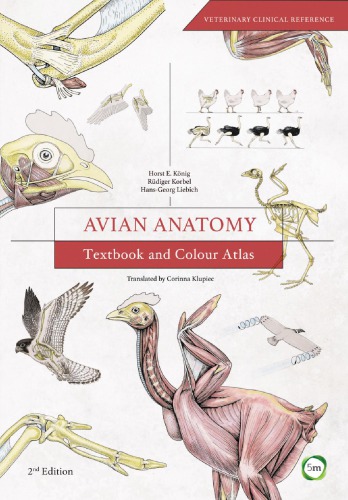
Avian Anatomy Textbook and Colour Atlas 2nd Edition by Horst Koenig, Ruediger Korbel 1910455601 9781910455609
Original price was: $50.00.$25.00Current price is: $25.00.
Avian Anatomy Textbook and Colour Atlas 2nd Edition by Horst Koenig, Ruediger Korbel – Ebook PDF Instant Download/Delivery: 1910455601, 9781910455609
Full dowload Avian Anatomy Textbook and Colour Atlas 2nd Edition after payment
Product details:
• ISBN 10:1910455601
• ISBN 13:9781910455609
• Author: Horst Koenig, Ruediger Korbel
Bringing together annotated images and anatomical terms, this reference book is a unique combination of a practical, clinically oriented textbook and pictorial atlas of avian anatomy. Containing very high quality photographs, including histological and radiographic images, and schematic diagrams, this edition focuses on ornamental birds and poultry. Among the various species examined are chickens, ducks, and geese, as well as budgerigars, psitaccines and many others. Wild bird species, such as the common buzzard and falcon, are included. Raptors are featured in a dedicated new chapter. Translated from Anatomie der Voegel, first published by Schattauer, this edition of Avian Anatomy is an ideal book for veterinary practitioners and students. *** “…a wealth of knowledge. Aside from anatomy, the book contains 7 chapters that are dedicated to clinically relevant topics, such as diagnostic imaging techniques, restraint and handling, and medication techniques. This book is an excellent reference for avian veterinarians, poultry specialists, veterinary students, and others interested in enhancing their knowledge of avian anatomy.” –Journal of the American Veterinary Medical Association, Vol. 252, No. 6, March 15, 2018[Subject: Veterinary Medicine, Avian Health]
Avian Anatomy Textbook and Colour Atlas 2nd Table of contents:
1 Introduction
— History of avian anatomy — Overview of the locomotion and anatomy of birds — Feathers — Skeletal adaptations for locomotion — Types of locomotion — Flight — Land– and water-based locomotion — Digestive system — Respiratory system — Urogenital apparatus — Avian egg and incubation period — Cardiovascular system — Brain and sense organs — Locomotor apparatus — Skeleton (systema skeletale) — Osteology (osteologia) — Structure of mature bone — Types of bone — Arthrology (syndesmologia)— Myology (myologia)
2 Head and trunk
— Skeleton of the head — Bones of the head — Cranium (ossa cranii) — Skeleton of the face (ossa faciei) — Joints of the head — Muscles of the head — Skeleton of the trunk— Vertebral column (columna vertebralis) — Cervical vertebrae (vertebrae cervicales) — Thoracic vertebrae (vertebrae thoracicae)— Synsacrum — Caudal vertebrae (vertebrae caudales) — Ribs (costae) — Sternum— Joints of the trunk— Joints of the vertebral column (juncturae columnae vertebralis) — Joints of the ribs (juncturae costarum) — Joints of the sternum (juncturae sterni) — Muscles of the trunk (musculi trunci)– Muscles of the vertebral column (musculi vertebrales)— Muscles of the thoracic and abdominal walls (musculi thoracis et abdominis) — Muscles of the tail (musculi caudae) — Clinical aspects
3 Thoracic limb (membrum thoracicum)
— Skeleton of the pectoral girdle and wing — Skeleton of the pectoral girdle (Ossa cinguli membri thoracici)
— Coracoid bone (os coracoideum) — Scapula — Clavicle (clavicula) — Skeleton of the wing (ossa alae) — Humerus
— Ulna and radius — Carpal bones (ossa carpi) — Metacarpal bones (ossa metacarpalia) Bones of the digits (ossa digitorum manus) — Joints of the pectoral girdle and wing — Joints of the pectoral girdle (juncturae cinguli membri thoracici) — Joints of the wing (juncturae alae) — Shoulder joint (articulatio humeri)
– Elbow joint (juncturae cubiti) — Joints of the carpus and manus (juncturae carpi et manus)— Muscles of the pectoral girdle and wing— Clinical aspects —
4 Pelvic limb (membrum pelvinum)
— Skeleton of the pelvic girdle and pelvic limb — Skeleton of the pelvic girdle (ossa cinguli membri pelvici) — Ilium (os ilium) — Ischium (os ischii) — Pubis (os pubis) — Skeleton of the pelvic limb (ossa membri pelvici) — Femur (os femoris) — Tibiotarsus
— Fibula — Tarsometatarsus — Digits — Joints of the pelvic girdle and pelvic limb — Joints of the pelvic girdle (juncturae cinguli membri pelvici)
— Joints of the pelvic limb (juncturae membri pelvici) — Knee joint (juncturae genus) — Intertarsal joint (articulatio intertarsalis)
— Joints of the metatarsal bones — Metatarsophalangeal joints (articulationes metatarsophalangeales)
— Interphalangeal joints (articulationes interphalangeales) — Muscles of the pelvic limb — Clinical aspects —
5 Body cavities
— Clinical aspects —
6 Digestive system (apparatus digestorius)
— Oral cavity (cavum oris) and pharynx — Beak, bill (rostrum)
— Roof of the oral cavity and pharynx (oropharynx) — Floor of the oral cavity
— Floor of the pharynx — Salivary glands (glandulae salivariae)
— Swallowing — Alimentary canal (canalis alimentarius) — Oesophagus — Crop (ingluvies) — Stomach (gaster)
— Proventriculus (glandular stomach, pars glandularis)
— Muscular stomach (ventriculus, pars muscularis) — Tunica mucosa gastris — Tunica muscularis gastris
— Intestine (intestinum) — Gut-associated lymphatic tissue (GALT)
— Small intestine (intestinum tenue) — Duodenum ejunum and ileum — Large intestine (intestinum crassum)
— Caeca — Rectum — Cloaca — Glands associated with the alimentary canal — Liver (hepar)
— Porta hepatis — Attachments of the liver — Gallbladder (vesica fellea) — Pancreas — Clinical aspects —
7 Respiratory system (apparatus respiratorius)
— Nasal cavity (cavum nasi) — Larynx — Trachea — Syrinx — Lung (pulmo) — Bronchial system and gas exchange
— Air sacs (sacci pneumatici, sacci aerophori) — Clinical aspects —
8 Urinary system (organa urinaria)
— Kidney (nephros, ren) — Structure of the kidney
— Structure of renal lobules — Renal corpuscle (corpusculum renis, Malpighian body) and nephron
— Tubules and collecting ducts — Juxtaglomerular apparatus
— Urine formation— Ureter — Clinical aspects —
9 Male genital organs (organa genitalia masculina)
— Testis (orchis) — Structure of the testis — Epididymis
— Deferent duct (ductus deferens) — Phallus (penis, phallus masculinus)
— Accessory structures of the phallus — Clinical aspects —
10 Female genital organs (organa genitalia feminina)
— Ovary (ovarium) — Oogenesis — Oviduct (oviductus) — Infundibulum — Magnum — Isthmus — Uterus (metra) — Vagina — Structure of the avian egg — Clinical aspects —
11 Cardiovascular system (systema cardiovasculare)
— Heart (cor) — Blood vessels of the heart — Conduction system of the heart — Pulmonary vessels — Systemic arteries — Brachiocephalic trunk (truncus brachiocephalicus) — Visceral branches of the descending aorta — Renal arteries — Arteries of the pelvic limb — Ischiadic artery — Arteries of the pelvic region — Systemic veins — Veins of the wing — Caudal vena cava (vena cava caudalis) — Hepatic portal system — Renal portal system — Clinical aspects —
12 Immune system and lymphatic organs (organa lymphopoetica)
Lymphatic vessels (systema lymphovasculare)
— Lymph heart (cor lymphaticum) — Avian lymph nodes and mural lymphoreticular formations — Avian lymph nodes — Mural lymphoreticular formations — Lymphatic organs (thymus, cloacal bursa and spleen) — Thymus — Cloacal bursa (bursa cloacalis, bursa Fabricii) — Spleen (lien, splen) — Clinical aspects —
13 Nervous system (systema nervosum)
— Central nervous system (systema nervosum centrale) — Spinal cord (medulla spinalis) — Brain (encephalon) — Nuclei of the medulla oblongata and pons — Metencephalon — Mesencephalon — Diencephalon — Telencephalon — Ventricles of the brain (ventriculi cerebri) — Meninges and meningeal blood vessels — Peripheral nervous system (systema nervosum periphericum) — Cranial nerves (nervi craniales) — Olfactory nerve (I) — Optic nerve (II) — Oculomotor nerve (III) — Trochlear nerve (IV) — Trigeminal nerve (V) — Abducent nerve (VI) — Facial nerve (VII) — Vestibulocochlear nerve (VIII) — Glossopharyngeal nerve (IX) — Vagus nerve (X) — Accessory nerve (XI) — Hypoglossal nerve (XII) — Spinal nerves (nervi spinales) — Brachial plexus (plexus brachialis) — Lumbosacral plexus (plexus lumbosacralis) — Lumbar plexus (plexus lumbalis) — Sacral plexus (plexus sacralis) — Pudendal plexus (plexus pudendus) — Caudal plexus (plexus caudae) — Autonomic nervous system (systema nervosum autonomicum) — Sympathetic system — Parasympathetic system — Clinical aspects —
14 Endocrine glands (glandulae endocrinae)
— Hypophysis, pituitary gland (glandula pituitaria) — Epiphysis, pineal gland (glandula pinealis) — Thyroid gland (glandula thyroidea) — Parathyroid gland (glandula parathyroidea) — Ultimobranchial body (glandula ultimobranchialis) — Adrenal gland (glandula adrenalis) — Pancreatic islets (insulae pancreaticae) — Gonads (testis, ovarium)
Clinical aspects —
15 The eye (organum visus)
— Orbit (orbita) — Eyeball, bulb (bulbus oculi) — Size, shape and position — Structure of the eyeball — Outer fibrous layer (tunica fibrosa or externa bulbi) — Sclera — Cornea — Middle vascular layer (tunica vasculosa or media bulbi, uvea) — Iris — Ciliary body (corpus ciliare) — Choroid (choroidea) — Inner layer (tunica interna bulbi, retina) — Optic nerve (nervus opticus) — Pecten (pecten oculi) — Internal structures of the eye — Lens — Anterior and posterior chambers (camera anterior and posterior bulbi) and aqueous humour (humor aquosus) — Vitreous body (corpus vitreum — camera vitrea bulbi) — Adnexa of the eye (organa oculi accessoria) — Extrinsic muscles of the eyeball — Eyelids (palpebrae) — Lacrimal apparatus (apparatus lacrimalis) — Innervation of the eye — Blood vessels of the eye — Clinical aspects — Ophthalmic examination — History — Observation and vision testing — General ophthalmic examination — Specialised ophthalmic examination —
16 The ear (organum vestibulocochleare)
— External ear (auris externa) — Middle ear (auris media) — Internal ear (auris interna) — Clinical aspects —
17 Common integument (integumentum commune)
— Featherless body regions — Skin glands — Accessory cutaneous structures — Patagia — Interdigital webs — Rhamphotheca and cere — Scales — Pads — Claws — Spurs — Feathered body regions — Feathers — Feather structure — Types of feathers — Feather replacement and moulting — Blood supply and innervation of the skin — Clinical aspects —
18 Clinical examination
— History and signalment — Observation — Physical examination — Further investigation — Laboratory techniques —
19 Imaging techniques
— Photography — Radiography — Principles of radiographic image acquisition — Positioning for the ventrodorsal view
People also search for Avian Anatomy Textbook and Colour Atlas 2nd:
avian anatomy textbook
avian anatomy textbook and colour atlas (second edition)
avian digestive system facts
avian anatomy and physiology
avian anatomy
You may also like…

Biology and other natural sciences - Ecology
Colour Atlas of Woody Plants and Trees 1st Edition by Bryan Bowes 0367473984 9780367473983
Uncategorized
Textbook of Oral Anatomy Physiology Histology and Tooth Morphology 2nd Edition Rajkumar
Medicine - Pediatrics
Color Atlas of Pediatric Anatomy Laparoscopy and Thoracoscopy 1st Edition Merrill Mchoney
Medicine - Clinical Medicine
Medicine - Surgery
Art Of Laparoscopic Surgery Textbook Atlas 4 Volumes 2nd Edition Palanivelu