Oxidative stress and redox signalling in Parkinson disease 1st Edition by Rodrigo Franco ISBN 9781788011914 1788011910
$50.00 Original price was: $50.00.$25.00Current price is: $25.00.
Oxidative stress and redox signalling in Parkinson disease 1st Edition by Rodrigo Franco – Ebook PDF Instant Download/Delivery: 9781788011914 ,1788011910
Full download Oxidative stress and redox signalling in Parkinson disease 1st Edition after payment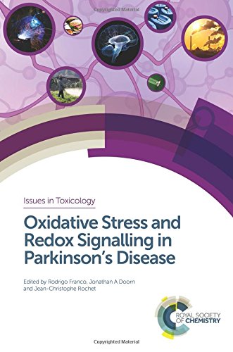
Product details:
ISBN 10: 1788011910
ISBN 13: 9781788011914
Author: Rodrigo Franco
This book provides a thorough review of the mechanisms by which oxidative stress and redox signalling mediate Parkinson’s Disease. Opening chapters bring readers up to speed on basic knowledge regarding oxidative stress and redox signalling, Parkinson’s Disease, and neurodegeneration before the latest advances in this field are explored in detail. Topics covered in the following chapters include the role of mitochondria, dopamine metabolism, metal homeostasis, inflammation, DNA-damage and thiol-signalling. The role of genetics and gene-environment interactions are also explored before final chapters discuss the identification of potential biomarkers for diagnosis and disease progression and the future of redox/antioxidant based therapeutics.
Written by recognized experts in the field, this book will be a valuable source of information for postgraduate students and academics, clinicians, toxicologists and risk assessment groups. Importantly, it presents the current research that might later lead to redox or antioxidant – based therapeutics for Parkinson’s disease.
Oxidative stress and redox signalling in Parkinson disease 1st Edition Table of contents:
CHAPTER 1 Etiology and Pathogenesis of Parkinson’s Disease
1.1 Introduction
1.2 Clinical Manifestations of Parkinson’s Disease
1.3 Neuropathology
1.3.1 Selective Vulnerability of the Nigrostriatal Dopamine Neuron
1.3.2 Mitochondrial Dysfunction in PD
1.3.3 Oxidative Stress
1.3.4 Dopamine Metabolism
1.3.5 Neuroinflammation
1.4 Genetics of Parkinson’s Disease
1.5 Environmental Exposures and the Risk of Parkinson’s Disease
1.5.1 Pesticides
1.5.2 Metals
1.5.3 Pathogens
1.6 Gene–Environment Interaction
1.7 Conclusions
Acknowledgements
References
CHAPTER 2 Oxidative Stress and Redox Signalling in the Parkinson’s Disease Brain
2.1 Introduction
2.2 Oxidative Stress and Antioxidant Systems
2.3 What Makes the Dopaminergic Neurons in the SNpc Vulnerable?
2.3.1 Cellular Organization of the SNpc
2.3.2 Redox Basis of the Vulnerability of SNpc DAergic Neurons
2.4 Conclusions and Perspectives
Acknowledgements
References
CHAPTER 3 Mitochondrial Dysfunction in Parkinson’s Disease
3.1 Reactive Oxygen Species (ROS)
3.1.1 Mitochondria and ROS Production
3.2 Parkinson’s Disease
3.3 Mitochondrial Dysfunction in PD
3.3.1 ETC Complex Deficiency in PD
3.3.2 Altered Mitochondrial Morphology
3.3.3 Mitochondrial Ca2+ Buffering in PD
3.3.4 PD-Related Genes and Mitochondrial Dysfunction
3.4 Mitochondrial Dysfunction in Toxicant-Induced PD
3.4.1 6-OHDA and MPTP: Classic Toxicant Models
3.4.2 Rotenone: A Case for Complex I Inhibition
3.4.3 Paraquat: Redox Cycling Agent
3.4.4 Maneb: A Role of Complex III in PD Pathogenesis
3.4.5 Other Environmental Toxins
3.5 Concluding Remarks
References
CHAPTER 4 Dopamine Metabolism and the Generation of a Reactive Aldehyde
4.1 The Life of Dopamine: Synthesis, Storage and Metabolism
4.2 3,4-Dihydroxyphenylacetaldehyde (DOPAL) and Biogenic Aldehydes Derived from Neurotransmitters
4.3 Generation of DOPAL and Biogenic Aldehydes at Aberrant Levels
4.3.1 Mechanisms for Elevation of DOPAL
4.3.2 Relevance of Altered Dopamine Metabolism/Trafficking to PD
4.4 Toxicity and Protein Reactivity of DOPAL and Biogenic Aldehydes
4.4.1 Mechanisms of Toxicity
4.4.2 Protein Reactivity and Targets
4.5 The Role of Biogenic Aldehydes in Disease
4.6 Summary
References
CHAPTER 5 Dopamine Oxidation and Parkinson’s Disease
5.1 Oxidative Stress and Susceptibility of Dopaminergic Neurons in Parkinson’s Disease
5.2 Dopamine Regulation and Metabolism
5.3 Pathological Dopamine Oxidation
5.3.1 Metabolism of Dopamine by Monoamine Oxidase
5.3.2 Oxidation of the Catechol Ring of Dopamine
5.3.3 Modification of Protein by Oxidized Dopamine
5.3.4 Mitochondrial Dysfunction and Dopamine Oxidation
5.4 The Role of Dopamine in Toxin-Induced Toxicity
5.5 Dopamine Toxicity
5.5.1 In vitro Exogenous Dopamine and l-DOPA Treatment
5.5.2 Exogenous Dopamine Administration in vivo
5.5.3 Dysregulation of Dopamine Handling in vivo
5.6 Dopamine and α-Synuclein
5.7 Dopamine Storage Disruption in PD
5.8 Summary and Conclusions
References
CHAPTER 6 Glutathione and Thiol Redox Signalling in Parkinson’s Disease
6.1 Introduction
6.2 Glutathione
6.3 Thiol Redox Signalling and Thiol–Disulfide Exchange
6.4 Cellular Reductases
6.4.1 Thioredoxins
6.4.2 Glutaredoxins
6.4.3 Peroxiredoxins
6.5 Glutathione Synthesis in the Brain
6.6 Glutathione and Models of Oxidative Stress in Dopaminergic Neurons
6.7 Glutathione Conjugating Enzymes
6.7.1 Glutathione Peroxidase
6.7.2 Glutathione S-Transferases
6.8 GSH and Transport in the Brain: Multidrug Resistance Proteins (MDRP) and the Blood–Brain Barrier (BBB)
6.9 Parkinson’s Disease Genetic Models and Redox Signalling
6.9.1 Glutathione-S-Transferase
6.9.2 DJ-1
6.9.3 PTEN-Induced Putative Kinase 1 (PINK1)
6.9.4 Parkin
6.10 Free Radicals as Messengers to Modulate Transcription Factors: Effects on Thiol Redox Regulation
6.11 Conclusions
References
CHAPTER 7 Neuroinflammation and Oxidative Stress in Models of Parkinson’s Disease and Protein-Misfolding Disorders
7.1 Introduction
7.2 Molecular Pathways Regulating Neuroinflammation in Glial Cells
7.2.1 Regulation of Inflammatory Genes in Glial Cells by NF-κB
7.2.2 Nuclear Regulation of NF-κB Function in Glial Cells
7.3 Neurotoxic Models of Parkinson’s Disease: Reactive Oxygen Species and Neuroinflammatory Mechanisms
7.3.1 1-Methyl-4-Phenyl-1,2,3,6-Tetrahydropyridine (MPTP)
7.3.2 6-Hydroxydopamine (6-OHDA)
7.3.3 Lipopolysaccharide (LPS)
7.4 Neuroinflammation and Protein Aggregation in PD and Protein-Misfolding Disorders
7.4.1 Neuroinflammation in Protein-Misfolding Disorders
7.4.2 Oxidative Stress in Protein-Misfolding Disorders
7.4.3 Unfolded Protein Response in Protein-Misfolding Disorders
7.5 Conclusions
References
CHAPTER 8 Redox Signalling in Dopaminergic Cell Death and Survival
8.1 Neuronal Degeneration in Parkinson’s Disease
8.1.1 Relatively Selective Dopaminergic Degeneration
8.1.2 Sources of Oxidative Stress and Selective Vulnerability
8.1.3 Increased Oxidative Stress and Selective Cell Death
8.2 Redox Signalling and Cell Survival/Death
8.3 Redox Regulation of DAergic Cell-Survival Pathways
8.3.1 Akt Structure and Function
8.3.2 Evidence of Akt1 Involvement in PD
8.3.3 Redox Regulation of Akt1 Activity
8.4 Redox Regulation of DAergic Cell-Death Pathways
8.4.1 Redox Regulation of MAP3K-ASK1
8.4.2 MAPKs – p38 and JNK
8.5 Conclusions
Acknowledgements
References
CHAPTER 9 Iron Metabolism in Parkinson’s Disease
9.1 Brain Iron Homeostasis
9.1.1 Brain Iron Transport and Distribution
9.1.2 Regulation of Brain Iron Homeostasis
9.2 Iron Metabolism and Parkinson’s Disease
9.2.1 Symptoms of Parkinson’s Disease
9.2.2 Iron Accumulation Accelerates the Symptomatology of Parkinson’s Disease
9.2.3 Mechanism of Iron Accumulation in the Substantia Nigra of Parkinson’s Disease Brains
9.2.4 Iron and the Aggregation of α-Synuclein
9.2.5 Iron, ROS and Apoptosis of Dopaminergic Neurons in the Substantia Nigra of Parkinson’s Disease
9.3 Iron-Related Therapeutic Approaches for PD
9.4 Conclusions
References
CHAPTER 10 Protein Oxidation, Quality-Control Mechanisms and Parkinson’s Disease
10.1 Introduction
10.2 Misfolded Protein Aggregation and Accumulation in PD
10.2.1 α-Synuclein: Mutations and Misfolding
10.3 Protein Quality-Control Mechanisms in PD
10.3.1 Protein Synthesis
10.3.2 Protein Folding, Unfolding and Disaggregation by Chaperones
10.3.3 Protein Degradation Pathways
10.3.4 Protein Quality Control in Organelles
10.4 Conclusions and Perspectives
Acknowledgements
References
CHAPTER 11 At the Intersection Between Mitochondrial Dysfunction and Lysosomal Autophagy: Role of PD-Related Neurotoxins and Gene Products
11.1 Introduction
11.2 Neuropathological Evidence for Mitochondrial Deficits and Autophagic Impairment in PD
11.2.1 Evidence of Mitochondrial Deficits in Postmortem PD Brains
11.2.2 Evidence of Autophagic Impairment in Postmortem PD Brains
11.3 Toxicological Evidence for Mitochondrial Deficits and Autophagic Impairment in PD
11.3.1 Rotenone
11.3.2 PQ and Maneb
11.3.3 MPTP or MPP+
11.3.4 6-OHDA
11.4 Genetic Evidence for Mitochondrial Deficits and Autophagic Impairment in PD
11.4.1 aSyn
11.4.2 Parkin/PINK1
11.4.3 DJ-1
11.4.4 ATP13A2
11.5 Interrelationships Between Autophagic Impairment and Mitochondrial Dysfunction
11.6 Summary and Future Directions
References
CHAPTER 12 Genes, Aging, and Parkinson’s Disease
12.1 Introduction
12.1.1 Do Genes Influence Lifespan?
12.1.2 Which Genes Influence Lifespan?
12.2 Apolipoprotein E
12.2.1 LDL Receptors
12.2.2 Effects of Polymorphisms on APOE Function
12.2.3 APOE and Parkinson’s Disease
12.2.4 APOE Oxidative Stress
12.3 FOXO3A and FOXO Family
12.3.1 FOXO3A Biological Functions
12.3.2 FOXO3A and Protein Homeostasis
12.3.3 FOXO3A and Parkinson’s Disease
12.4 Role of Other Genes Emerged from Animal Studies in Aging
12.4.1 Are Aging-Modifying Genes Discovered in Laboratory Animals Relevant for PD?
References
CHAPTER 13 Biomarkers of Oxidative Stress in Parkinson’s Disease
13.1 Biomarkers
13.2 Oxidative Stress
13.3 Candidate Biomarkers for ROS-Induced Stress
13.3.1 Halogenation
13.3.2 Methionine Oxidation
13.3.3 Glycation and Carbonylation
13.4 Candidate Biomarkers for RNS-Induced Stress
13.4.1 Nitration
13.4.2 S-nitros(yl)ation
13.5 Conclusions
References
CHAPTER 14 Dietary Anti-, Pro-Oxidants in the Etiology of Parkinson’s Disease
14.1 Introduction
14.2 Heterocyclic Amines
14.3 Polyphenolic Compounds
14.3.1 Flavonoids
14.3.2 Non-Flavonoids
14.4 Vitamins
14.4.1 Vitamin A
14.4.2 Vitamin B
14.4.3 Vitamin C
14.4.4 Vitamin D
14.4.5 Vitamin E
14.5 Summary
References
Subject Index
People also search for Oxidative stress and redox signalling in Parkinson disease 1st Edition:
autophagy mitochondria and oxidative stress cross talk and redox signalling
ros function in redox signaling and oxidative stress
oxidative stress simple explanation
oxidative stress simplified
Tags:
Rodrigo Franco,Oxidative stress,signalling,Parkinson disease
You may also like…
Medicine
Aging : Oxidative Stress and Dietary Antioxidants 2nd edition Victor 0128188111 9780128188118
Science (General)
Measuring Oxidants and Oxidative Stress in Biological Systems Lawrence J. Berliner
Biology and other natural sciences
Science (General)
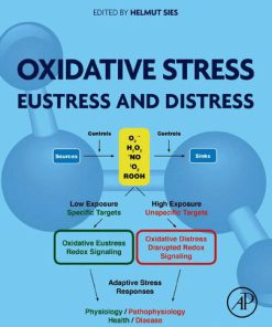
Oxidative Stress: Eustress and Distress 1st Edition Helmut Sies (Editor)
Medicine - Natural Medicine
Cancer: Oxidative Stress and Dietary Antioxidants 2nd Edition Vinood B. Patel
Medicine
Aging: Oxidative Stress and Dietary Antioxidants 2nd Edition Victor 0128188111 9780128188118
Biology and other natural sciences


