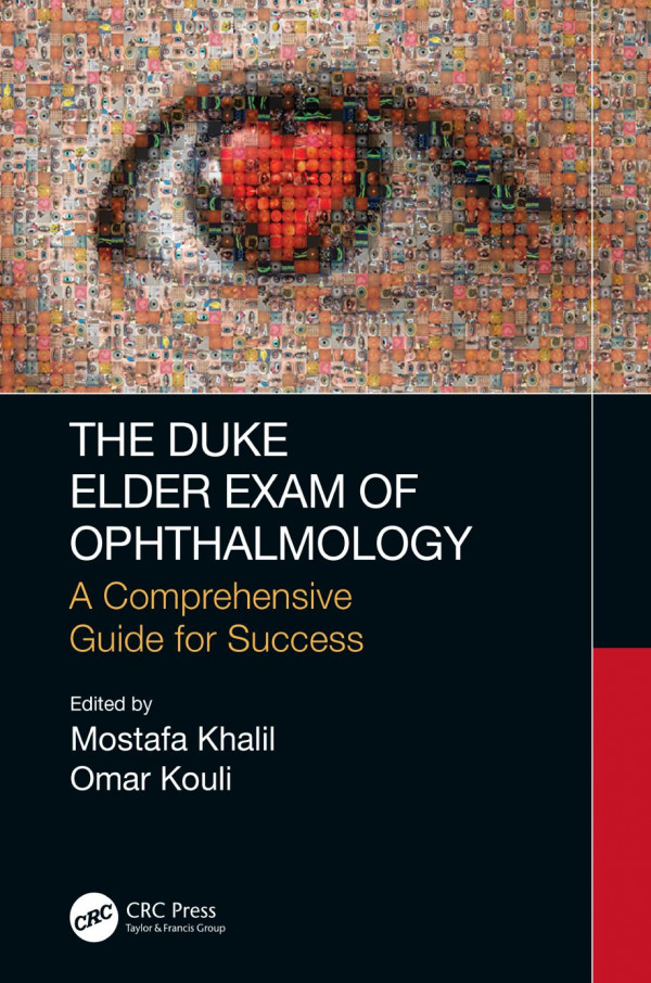
The Duke Elder Exam of Ophthalmology A Comprehensive Guide for Success 1st Edition by Mostafa Khalil, Omar Kouli 9781000020588 1000020584
$50.00 Original price was: $50.00.$25.00Current price is: $25.00.
The Duke Elder Exam of Ophthalmology A Comprehensive Guide for Success 1st Edition by Mostafa Khalil, Omar Kouli – Ebook PDF Instant Download/Delivery: 9781000020588, 1000020584
Full download The Duke Elder Exam of Ophthalmology A Comprehensive Guide for Success 1st Edition after payment
Product details:
• ISBN 10:1000020584
• ISBN 13:781000020588
• Author:Mostafa Khalil, Omar Kouli
The Duke Elder Exam of Ophthalmology
A Comprehensive Guide for Success
The Duke Elder Exam of Ophthalmology – A Comprehensive Guide for Success is an indispensable resource for any student wishing to achieve the highest mark on the Duke Elder Exam and receive a prize. With expert knowledge of students and doctors that have scored high on the exam, along with the supervision of well-regarded ophthalmologists and trainees, we believe this is the only resource you will need to achieve a high score on the exam. Key Features In-depth coverage of the Duke Elder Curriculum including the basic sciences, anatomy, optics and all subspecialties of ophthalmology Full colour and easy to read with clinical photographs and diagrams to aid in the understanding of key topics 180 SBAs, which accurately reflect the format and difficulty of the exam
The Duke Elder Exam of Ophthalmology A Comprehensive Guide for Success 1st Table of contents:
Preface
Acknowledgements
Editors
Contributors
Exam format
Author tips
1. Basic science, investigations and lasers
1.1 Embryologic origins of eye structures
1.2 Genetics
1.3 Microbiology
1.4 Immunology
1.5 Drug mechanisms and side effects
1.6 Investigations
1.7 Lasers
1.8 Miscellaneous
References
2. Optics and refractive errors
2.1 How do we see?
2.2 Myopia
2.3 Hypermetropia
2.4 Concepts relating to myopia and hypermetropia
2.5 Astigmatism
2.6 Concepts relating to astigmatism
2.7 Presbyopia
2.8 Concepts relating to presbyopia
2.9 Squints
2.10 Concepts relating to prisms
2.11 Measuring visual acuity
2.12 Optics of slit lamps
2.13 Optics of direct ophthalmoscopy
2.14 Optics of indirect ophthalmoscopy
References
3. Eyelids
3.1 Anatomy and physiology
3.2 Chalazion
3.3 Port wine stain (naevus flammeus)
3.4 Basal cell carcinoma
3.5 Squamous cell carcinoma
3.6 Sebaceous gland carcinoma
3.7 Trichiasis
3.8 Distichiasis
3.9 Blepharitis
3.10 Ptosis
3.11 Pseudoptosis
3.12 Floppy eyelid syndrome
3.13 Lid retraction
3.14 Lid coloboma
3.15 Hordeolum (stye)
4. Lacrimal system and dry eyes
4.1 Anatomy and physiology
4.2 Acquired obstruction
4.3 Congenital nasolacrimal duct obstruction
4.4 Canaliculitis
4.5 Dacryoadenitis
4.6 Dacryocystitis
4.7 Pleomorphic adenoma of the lacrimal gland
4.8 Lacrimal gland carcinoma
4.9 Sjögren syndrome
4.10 Xerophthalmia
5. Orbit
5.1 Anatomy
5.2 Thyroid eye disease
5.3 Orbital cellulitis and preseptal cellulitis
5.4 Orbital mucormycosis
5.5 Rhabdomyosarcoma
5.6 Neuroblastoma
5.7 Lymphangioma
5.8 Optic nerve glioma versus optic nerve sheath meningioma
5.9 Carotid-cavernous fistula: Direct versus indirect
5.10 Cavernous haemangioma
5.11 Capillary haemangioma
5.12 Cavernous sinus thrombosis
References
6. Strabismus
6.1 Anatomy and physiology
6.2 Important definitions
6.3 Esotropia
6.4 Exotropia
6.5 Microtropia
6.6 Duane retraction syndrome
6.7 Brown syndrome
6.8 Principles of strabismus surgery
Reference
7. Neuro-ophthalmology
7.1 Anatomy
7.2 Optic neuropathy
7.3 Pupils
7.4 Cranial nerves
7.5 Gaze abnormalities
7.6 Nystagmus
7.7 Myasthenia gravis
7.8 Myotonic dystrophy
7.9 Kearns-Sayre syndrome
7.10 Miller Fisher syndrome
7.11 Neurofibromatosis type 1
7.12 Neurofibromatosis type 2
7.13 Tuberous sclerosis
7.14 Benign essential blepharospasm
References
8. Cornea
8.1 Anatomy
8.2 Bacterial keratitis
8.3 Fungal keratitis
8.4 Acanthamoeba keratitis
8.5 Herpes simplex keratitis
8.6 Herpes zoster ophthalmicus
8.7 Interstitial keratitis (IK)
8.8 Marginal keratitis
8.9 Peripheral ulcerative keratitis
8.10 Ocular rosacea
8.11 Filamentary keratitis
8.12 Keratoconus
8.13 Microphthalmia
8.14 Wilson disease
8.15 Band keratopathy
8.16 Corneal dystrophies
9. Conjunctiva
9.1 Anatomy
9.2 Acute bacterial conjunctivitis
9.3 Adult inclusion (chlamydial) conjunctivitis
9.4 Trachoma
9.5 Ophthalmia neonatorum
9.6 Viral conjunctivitis
9.7 Allergic conjunctivitis
9.8 Ocular mucous membrane pemphigoid
9.9 Superior limbic keratoconjunctivitis
9.10 Parinaud oculoglandular syndrome
9.11 Pinguecula
9.12 Pterygium
References
10. Sclera
10.1 Anatomy
10.2 Episcleritis
10.3 Scleritis
10.4 Blue sclera
11. Lens and cataracts
11.1 Anatomy and physiology
11.2 Cataract maturity
11.3 Age-related cataracts
11.4 Acquired cataracts and their associations
11.5 Management
11.6 Complications of surgery
11.7 Congenital cataracts
11.8 Lenticonus
11.9 Ectopia lentis
References
12. Glaucoma
12.1 Anatomy and physiology
12.2 Ocular hypertension
12.3 Primary open-angle glaucoma
12.4 Normal-tension glaucoma
12.5 Primary angle-closure glaucoma
12.6 Neovascular glaucoma (NVG)
12.7 Pigment dispersion syndrome
12.8 Pseudoexfoliation syndrome
12.9 Posner-Schlossman syndrome
12.10 Phacolytic glaucoma
12.11 Phacomorphic glaucoma
12.12 Red cell glaucoma
12.13 Ghost cell glaucoma
12.14 Angle recession glaucoma
12.15 Sturge-Weber syndrome
12.16 Primary congenital glaucoma
References
13. Uveitis
13.1 Anatomy
13.2 Uveitis
13.3 Sarcoidosis
13.4 Tuberculosis
13.5 Sympathetic ophthalmia
13.6 Vogt-Koyanagi-Harada syndrome
13.7 Birdshot choroidopathy
13.8 Fuchs’ heterochromic cyclitis
13.9 Tubulointerstitial nephritis and uveitis
13.10 Juvenile idiopathic arthritis
13.11 Reiter syndrome
13.12 Behçet disease
13.13 Kawasaki disease
13.14 Onchocerciasis
13.15 Toxocariasis
13.16 AIDS-related conditions
13.17 Toxoplasmosis
13.18 Presumed ocular histoplasmosis syndrome
13.19 Cytomegalovirus retinitis
13.20 Progressive outer retinal necrosis
13.21 Acute retinal necrosis
14. Medical retina
14.1 Anatomy and physiology
14.2 Diabetic retinopathy and maculopathy
14.3 Hypertensive retinopathy
14.4 Retinal vein occlusion
14.5 Retinal artery occlusion
14.6 Ocular ischaemic syndrome
14.7 Sickle cell retinopathy
14.8 Age-related macular degeneration
14.9 Choroidal neovascularization (CNV)
14.10 Degenerative myopia
14.11 Angioid streaks
14.12 Cystoid macular oedema
14.13 Central serous chorioretinopathy
14.14 Eales disease
14.15 Best disease
14.16 Stargardt disease
14.17 Lebers congenital amaurosis
14.18 Albinism
14.19 Retinitis pigmentosa
14.20 Retinitis pigmentosa-related conditions
14.21 Causes of leukocoria
14.22 Coats’ disease
14.23 Von Hippel-Lindau
14.24 Choroidal melanoma
References
15. Vitreoretinal
15.1 Peripheral retinal degeneration
15.2 Posterior vitreous detachment
15.3 Retinal breaks
15.4 Retinal detachment
15.5 Vitreous haemorrhage
15.6 Choroidal detachment
15.7 Choroidal rupture
15.8 Uveal effusion syndrome
15.9 Epiretinal membrane
15.10 Macular hole
15.11 Hereditary vitreoretinal degenerations
References
16. Ocular trauma
16.1 Lefort fractures
16.2 Orbital floor fracture
16.3 Intraocular foreign bodies
16.4 Chemical injury
16.5 Shaken baby syndrome
16.6 Purtscher retinopathy
Questions & Answers
Paper 1: Questions
Paper 1: Answers
Paper 2: Questions
Paper 2: Answers
Index
Read Less
People also search for The Duke Elder Exam of Ophthalmology A Comprehensive Guide for Success 1st:
the duke elder exam of ophthalmology
how to prepare for duke elder exam
duke elder exam results
duke eye center near me
duke elder system of ophthalmology
You may also like…
Business & Economics - Project Management
Achieve CAPM Exam Success A Concise Study Guide for New Project Manager 3rd Edition Diane Altwies
Computers - Networking
Storage Design and Implementation in vSphere 6 A Technology Deep Dive 2nd Edition Mostafa Khalil
Business & Economics - Management & Leadership
Computers - Computer Certification & Training


