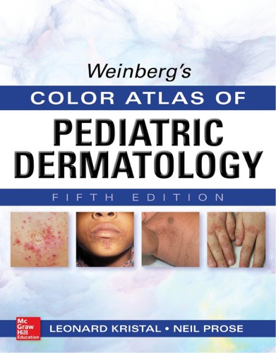
Weinberg Color Atlas of Pediatric Dermatology Fifth Edition by Leonard Kristal, Neil Prose 9780071792264 0071792260
$50.00 Original price was: $50.00.$25.00Current price is: $25.00.
Weinberg Color Atlas of Pediatric Dermatology Fifth Edition by Leonard Kristal, Neil Prose – Ebook PDF Instant Download/Delivery: 9780071792264, 0071792260
Full dowload Weinberg Color Atlas of Pediatric Dermatology Fifth Edition after payment
Product details:
• ISBN 10:0071792260
• ISBN 13:978007179226
• Author:Leonard Kristal, Neil Prose
Weinberg’s Color Atlas of Pediatric Dermatology, Fifth Edition
An unsurpassed visual archive of pediatric dermatology — with more than 300 brand-new color images! Color Atlas of Pediatric Dermatology contains a collection of more than 900 full-color images that provide invaluable guidance for the diagnosis of both common and rare skin disorders of neonates, infants, and children. For each condition reviewed in the text, one or more clinical photographs are supplied, making differential diagnosis faster, easier, and more accurate. A concise, yet thorough overview of important clinical features, etiology, prognosis, and the most current therapeutic approaches is included for each disorder illustrated. Every page reflects the book’s magnificent atlas format, with photographs linked to brief text that authoritatively describes the condition illustrated in each image, along with, in most cases, a one-sentence summary of suggested treatment protocols. New treatment modalities are included when applicable, reflecting the very latest clinical findings and treatment guidelines Revised and refreshed legends throughout the text to highlight the latest thinking in pediatric dermatology practice
Weinberg Color Atlas of Pediatric Dermatology Fifth Table of contents:
Section 1 Benign Neonatal Dermatoses
Figures 1-1–1-4 Erythema toxicum neonatorum
1-5–1-8 Transient neonatal pustular melanosis
Section 2 Milia, Miliaria, and Pustular and Acneiform Disorders
Figures 2-1, 2-2 Milia
2-3 Eosinophilic pustular folliculitis of infancy
2-4–2-7 Infantile acropustulosis
2-8, 2-9 Miliaria crystallina
2-10 Miliaria rubra (prickly heat)
2-11, 2-12 Fox-Fordyce disease (apocrine miliaria)
2-13, 2-14 Neonatal cephalic pustulosis
2-15–2-17 Neonatal and infantile acne
2-18 Infantile cystic acne
2-19–2-24 Acne vulgaris
2-25, 2-26 Cystic acne
2-27 Acne conglobata
2-28, 2-29 Scarring following acne
2-30 Acne and precocious puberty from a pinealoma
2-31–2-33 Steroid acne (dexamethasone)
2-34–2-36 Hidradenitis suppurativa
2-37 Rosacea
2-38, 2-39 Periorificial dermatitis
2-40, 2-41 Periorificial granulomatous dermatitis
2-42 Periorificial dermatitis
2-43, 2-44 Dissecting cellulitis of the scalp
Section 3 Bacterial Infections
Figures 3-1, 3-2 Impetigo
3-3, 3-4 Bullous impetigo
3-5 Impetiginization
3-6 Ecthyma
3-7–3-11 Staphylococcal scalded skin syndrome
3-12 Chancriform pyoderma
3-13–3-15 Folliculitis
3-16 Hot tub folliculitis
3-17, 3-18 Furuncle
3-19–3-22 Streptococcal intertrigo
3-23 Perianal streptococcal disease
3-24 Blistering distal dactylitis
3-25–3-30 Scarlet fever
3-31 Erysipelas
3-32 Invasive group A streptococcal disease
3-33, 3-34 Cat-scratch disease
3-35, 3-36 Erythrasma
3-37, 3-38 Verruga peruana (Carrion disease)
3-39 Pitted keratolysis
3-40 Actinomycosis
3-41, 3-42 Cutaneous effects of Pseudomonas sepsis
3-43, 3-44 Cutaneous effects of meningococcemia
3-45, 3-46 Cutaneous effects of gonococcemia
Section 4 Spirochetal, Protozoal, Mycobacterial, and Rickettsial Diseases
Figures 4-1, 4-2 Congenital syphilis
4-3–4-6 Acquired syphilis
4-7 Yaws
4-8–4-11 Erythema migrans (Lyme disease)
4-12, 4-13 Lepromatous leprosy
4-14, 4-15 Tuberculoid leprosy
4-16, 4-17 Dimorphous leprosy
4-18 Primary complex of tuberculosis in the skin
4-19 Scrofuloderma
4-20, 4-21 Lupus vulgaris
4-22, 4-23 Tuberculosis cutis verrucosa
4-24 Lichen scrofulosorum
4-25 Papulonecrotic tuberculid
4-26–4-28 Infection with atypical mycobacteria
4-29 Amebiasis cutis
4-30, 4-31 Leishmaniasis
4-32, 4-33 Rickettsialpox
4-34, 4-35 Rocky Mountain spotted fever
Section 5 Viral Diseases
Figures 5-1–5-8 Molluscum contagiosum
5-9, 5-10 Molluscum contagiosum dermatitis
5-11, 5-12 Molluscum contagiosum id reaction
5-13–5-20 Verruca vulgaris
5-21, 5-22 Plantar warts
5-23, 5-24 Filiform warts
5-25, 5-26 Verruca plana
5-27, 5-28 Condyloma acuminatum
5-29 Epidermodysplasia verruciformis
5-30, 5-31 Neonatal herpes simplex
5-32 Herpes simplex
5-33 Herpetic whitlow
5-34 Herpetic gingivostomatis
5-35 Recurrent herpes simplex
5-36–5-38 Herpes simplex, recurrent
5-39 Genital herpes simplex
5-40, 5-41 Eczema herpeticum (Kaposi varicelliform eruption)
5-42–5-48 Varicella
5-49 Congenital varicella syndrome
5-50, 5-51 Neonatal varicella
5-52–5-55 Herpes zoster
5-56–5-58 Hand-foot-mouth disease
5-59–5-62 Atypical hand-foot-mouth disease
5-63–5-65 Complications of vaccinia
5-66 Eczema vaccinatum
5-67, 5-68 Exanthem subitum (roseola)
5-69, 5-70 Unilateral laterothoracic exanthem
5-71–5-74 Papular acrodermatitis of childhood (Gianotti-Crosti syndrome)
5-75 Congenital cytomegalovirus infection
5-76 Congenital rubella
5-77, 5-78 Rubella
5-79–5-81 Measles
5-82 Atypical measles
5-83–5-86 Erythema infectiosum (fifth disease)
5-87, 5-88 Papular purpuric sock and glove syndrome
Section 6 Superficial Fungal Infections
Figures 6-1–6-3 Tinea corporis
6-4 Tinea corporis-vesicular
6-5, 6-6 Tinea corporis
6-7–6-10 Tinea corporis (faciei)
6-11, 6-12 Neonatal tinea
6-13, 6-14 Tinea incognito
6-15–6-19 Tinea capitis
6-20 Kerion
6-21 Tinea capitis
6-22 Id reaction to tinea
6-23, 6-24 Tinea corporis secondary to scalp infection
6-25, 6-26 Tinea cruris
6-27–6-30 Tinea pedis
6-31, 6-32 Onychomycosis
6-33 White superficial onychomycosis
6-34 Tinea manuum
6-35–6-40 Tinea versicolor
6-41, 6-42 Tinea nigra
6-43 Tinea imbricata
6-44 Favus
6-45–6-48 Congenital cutaneous candidiasis
6-49, 6-50 Candidiasis (moniliasis)
6-51, 6-52 Chronic mucocutaneous candidiasis
Section 7 Deep Fungal Infections
Figures 7-1 Chromoblastomycosis
7-2, 7-3 Coccidioidomycosis
7-4–7-7 Sporotrichosis
7-8, 7-9 Mycetoma
Section 8 Bites and Infestations
Figures 8-1–8-7 Insect “bites”
8-8 Dermatitis caused by the common carpet beetle
8-9 Spider bites
8-10, 8-11 Papular urticaria
8-12 Ticks
8-13 Tick “bite”
8-14 Tick bite granuloma
8-15–8-25 Scabies
8-26 Postscabetic acropustulosis
8-27 Scabies
8-28–8-31 Pediculosis capitis (head lice)
8-32 Pediculosis corporis
8-33 Pediculosis pubis
8-34 Macula ceruleae
8-35, 8-36 Myiasis
8-37 Caterpillar dermatitis
8-38 Dermatitis caused by blister beetles
8-39 Seabather’s eruption
8-40 Swimmer’s itch
8-41 The sting of the Portuguese man-of-war
8-42 The effect of contact with a sea urchin
8-43–8-46 Larva migrans (creeping eruption)
8-47–8-48 Onchocerciasis
Section 9 Atopic Dermatitis
Figures 9-1–9-17 Atopic dermatitis
9-18, 9-19 Juvenile plantar dermatosis
9-20, 9-21 Frictional lichenoid dermatitis
9-22 Atopic dermatitis with follicular accentuation
9-23 Lichen spinulosus
9-24, 9-25 Pityriasis alba
9-26, 9-27 Pompholyx (dyshidrotic eczema)
Section 10 Allergic and Irritant Contact Dermatitis
Figure 10-1–10-4 Allergic contact dermatitis (Poison ivy)
10-5 Allergic contact dermatitis (wet wipes)
10-6 Allergic contact dermatitis (disposable diapers)
10-7 Allergic contact dermatitis (mango)
10-8 Allergic contact dermatitis (neomycin)
10-9 Allergic contact dermatitis (shoes)
10-10 Allergic contact dermatitis
10-11–10-14 Nickel contact dermatitis
10-15, 10-16 Allergic contact dermatitis (Reaction to temporary tattoo)
10-17 Irritant diaper dermatitis
10-18, 10-19 Erosive diaper dermatitis (dermatitis of Jacquet)
10-20 Pseudoverrucous papules and nodules
10-21, 10-22 Lip licking dermatitis
10-23 Shin guard dermatitis
Section 11 Photodermatoses
Figure 11-1–11-4 Polymorphous light eruption
11-5 Photoallergic dermatitis
11-6 Photoxic dermatitis
11-7, 11-8 Erythropoietic protoporphyria
11-9, 11-10 NSAID-induced pseudoporphyria
11-11 Actinic prurigo
11-12 Actinic prurigo cheilitis
11-13, 11-14 Hydroa vacciniforme
11-15, 11-16 Juvenile spring eruption
11-17 Photodermatitis (berloque dermatitis)
11-18–11-20 Phytophotodermatitis
Section 12 Papulosquamous Diseases
Figure 12-1–12-8 Seborrheic dermatitis
12-9 Tinea amiantacea
12-10–12-27 Psoriasis
12-28–12-31 Pustular psoriasis
12-32–12-37 Pityriasis rubra pilaris
12-38–12-41 Pityriasis rosea
12-42–12-45 Pityriasis lichenoides chronica
12-46–12-51 Pityriasis lichenoides et varioliformis acuta (PLEVA, Mucha-Habermann disease)
12-52–12-55 Lichen nitidus
12-56–12-63 Lichen striatus
12-64–12-67 Follicular mucinosis
12-68, 12-69 Porokeratosis
12-70 Porokeratosis (porokeratosis of Mibelli)
12-71 Linear porokeratosis
12-72, 12-73 Elastosis perforans serpiginosa
12-74 Perforating folliculitis
12-75 Reactive perforating collagenosis
12-76–12-85 Lichen planus
12-86 Actinic lichen planus
12-87 Annular lichen planus
12-88, 12-89 Lichen planopilaris (follicular lichen planus)
Section 13 Nutritional, Metabolic, and Endocrine Diseases
Figure 13-1–13-6 Acrodermatitis enteropathica
13-7–13-9 Kwashiorkor
13-10 Marasmus
13-11, 13-12 Pellagra
13-13–13-15 Lipoid proteinosis
13-16 Hurler syndrome
13-17–13-21 Xanthomatosis
13-22, 13-23 Calcinosis cutis
13-24 Progressive osseous heteroplasia
13-25–13-28 Acanthosis nigricans
Section 14 Genodermatoses
Figure 14-1–14-3 Pseudoxanthoma elasticum
14-4 Cutis laxa
14-5–14-11 Ehlers-Danlos syndrome
14-12–14-16 Focal dermal hypoplasia (Goltz syndrome)
14-17–14-26 Incontinentia pigmenti
14-27 Wiskott-Aldrich syndrome
14-28, 14-29 Ataxia telangiectasia
14-30 Bloom syndrome
14-31 Rothmund-Thomson syndrome (Poikiloderma congenitale)
14-32, 14-33 Cockayne syndrome
14-34, 14-35 Hypohidrotic ectodermal dysplasia
14-36, 14-37 Clouston syndrome
14-38–14-41 Hay-Well syndrome (AEC)
14-42–14-45 Pachyonychia congenita
14-46–14-49 Dyskeratosis congenita
14-50–14-53 Neurofibromatosis
14-54–14-57 Neurofibromatosis (von Recklinghausen disease)
14-58, 14-59 Neurofibromatosis type I (von Recklinghausen disease)
14-60 Neurofibromatosis type I
14-61 Neurofibromatosis type I (Lisch nodule)
14-62, 14-63 Multiple endocrine neoplasia type 2
14-64–14-70 Tuberous sclerosis
14-71 Buschke-Ollendorf syndrome
14-72–14-74 Basal cell nevus syndrome
14-75–14-77 Xeroderma pigmentosum
Section 15 Ichthyoses and Disorders of Keratinization
Figure 15-1–15-4 Ichthyosis vulgaris
15-5 Harlequin-type ichthyosis
15-6–15-8 Collodion baby
15-9–15-12 Lamellar ichthyosis
15-13, 15-14 Lamellar icthyosis (cont’d.)
15-15, 15-16 Lamellar ichthyosis
15-17–15-20 Recessive X-linked ichthyosis
15-21–15-27 Epidermolytic ichthyosis (formerly epidermolytic hyperkeratosis)
15-28–15-30 Nonbullous congenital ichthyosiform erythroderma
15-31 Erythrokeratoderma variabilis
15-32–15-34 Progressive symmetric erythrokeratoderma
15-35–15-40 Netherton syndrome
15-41, 15-42 Palmoplantar keratoderma
15-43, 15-44 Conradi-Hünermann syndrome
15-45, 15-46 Sjögren-Larsson syndrome
15-47–15-50 Linear epidermal nevus
15-51, 15-52 Epidermal nevus syndrome
15-53, 15-54 Inflammatory linear verrucous epidermal nevus (ILVEN)
15-55–15-57 Darier disease (keratosis follicularis)
15-58 Acrokeratosis verruciformis
15-59, 15-60 KID syndrome
15-61, 15-63 Keratosis pilaris
15-64 Keratosis pilaris rubra faciei
Section 16 Urticarial, Purpuric, and Vascular Reactions
Figure 16-1–16-4 Urticaria
16-5, 16-6 Physical urticarias
16-7–16-12 Erythema multiforme
16-13–16-20 Stevens-Johnson syndrome/toxic epidermal necrolysis
16-21–16-23 Urticaria multiforme
16-24 Sweet syndrome (acute febrile neutrophilic dermatosis)
16-25, 16-26 Erythema annulare centrifugum
16-27, 16-28 Erythema elevatum diutinum
16-29–16-32 Progressive pigmented purpura (Schamberg disease)
16-33, 16-34 Acute hemorrhagic edema of infancy
16-35, 16-36 Traumatic purpura
16-37–16-40 Henoch-Schönlein purpura
16-41, 16-42 Purpura fulminans
16-43, 16-44 Aphthous stomatitis
16-45–16-47 Behçet syndrome
16-48, 16-49 Erythema ab igne
Section 17 Bullous, Pustular, and Ulcerating Diseases
Figure 17-1–17-4 Pemphigus vulgaris
17-5 Pemphigus vegetans
17-6 Familial benign chronic pemphigus (Hailey-Hailey disease)
17-7, 17-8 Subcorneal pustulosis (Sneddon-Wilkinson disease)
17-9–17-12 Pemphigus foliaceus
17-13, 17-14 Bullous pemphigoid
17-15 Vulvar pemphigoid
17-16–17-18 Epidermolysis bullosa simplex (EBS)
17-19 Epidermolysis bullosa simplex (Dowling-Meara)
17-20–17-22 Epidermolysis bullosa, junctional type (JEB)
17-23–17-25 Epidermolysis bullosa recessive dystrophic type (RDEB)
17-26 Epidermolysis bullosa dominant dystrophic type, generalized (DDEB)
17-27–17-29 Dystrophic epidermolysis bullosa, dominant type (DDEB)
17-30 Epidermolysis bullosa with congenital absence of skin
17-31–17-33 Dermatitis herpetiformis
17-34–17-37 Linear Ig A dermatosis (chronic bullous dermatosis of childhood)
Section 18 Cutaneous Manifestations of HIV Disease
Figure 18-1 Cutaneous manifestations of HIV infection (chronic varicella zoster infection)
18-2 Cutaneous manifestations of HIV infection (herpes zoster infection)
18-3 Cutaneous manifestations of HIV infection (scarring from herpes zoster)
18-4 Cutaneous manifestations of HIV infection (candidal paronychias and nail dystrophy)
18-5 Cutaneous manifestations of HIV infection (drug eruption)
18-6 Cutaneous manifestations of HIV infection (molluscum contagiosum)
18-7 Cutaneous manifestations of HIV infection (chronic herpetic gingivostomatitis)
18-8 Cutaneous manifestations of HIV infection (seborrheic dermatitis)
18-9 Cutaneous manifestations of HIV infection (condylomata acuminata)
18-10 Cutaneous manifestations of HIV infection (widespread flat warts)
18-11, 18-12 Cutaneous manifestations of HIV infection (psoriasis)
Section 19 Cutaneous Manifestations of Systemic Disease
Figure 19-1–19-6 Systemic lupus erythematosus
19-7 Systemic lupus erythematosus vasculitis
19-8–19-11 Neonatal lupus erythematosus
19-12–19-15 Discoid lupus erythematosus
19-16–19-25 Dermatomyositis
19-26, 19-27 Scleroderma (progressive systemic sclerosis)
19-28 Cutaneous expression of rheumatic fever
19-29 Cutaneous expression of rheumatoid arthritis
19-30, 19-31 Cutaneous expressions of polyarteritis nodosa
19-32, 19-33 Sarcoidosis
19-34, 19-35 Pernio
19-36, 19-37 Cutaneous manifestations of Crohn disease
19-38, 19-39 Pyoderma gangrenosum
19-40–19-45 Kawasaki disease
19-46, 19-47 Necrobiosis lipoidica
Section 20 Disorders of the Dermis (Infiltrates, Atrophies, and Nodules)
Figure 20-1, 20-2 Morphea
20-3–20-5 Morphea (linear)
20-6 Morphea (linear, en coup de sabre)
20-7, 20-8 Morphea
20-9, 20-10 Generalized morphea
20-11 Atrophoderma (Pasini-Pierini)
20-12–20-15 Lichen sclerosus et atrophicus
20-16 Lichen sclerosus et atrophicus (Balanitis xerotica obliterans)
20-17, 20-18 Anetoderma (macular atrophy)
20-19–20-21 Striae distensae
20-22, 20-23 Connective tissue nevus
20-24–20-26 Granuloma annulare
20-27, 20-28 Subcutaneous granuloma annulare
20-29 Eruptive granuloma annulare
20-30–20-37 Juvenile xanthogranuloma
20-38, 20-39 Dermatofibroma
20-40, 20-41 Granular cell tumor
20-42, 20-43 Benign cephalic histiocytosis
20-44–20-46 Keloids
20-47 Fibrous hamartoma of infancy
20-48–20-51 Digital fibrous tumor of childhood
20-52, 20-53 Infantile myofibromatosis
20-54, 20-55 Mastocytoma
20-56–20-59 Mastocytosis
20-60, 20-61 Bullous mastocytosis (Diffuse cutaneous mastocytosis)
20-62 Nevus lipomatosus superficialis
20-63 Leiomyoma
20-64, 20-65 Smooth muscle and pilar hamartoma
20-66–20-68 Lymphomatoid papulosis
Section 21 Drug Eruptions
Figure 21-1–21-3 Drug eruptions
21-4 Urticaria multiforme
21-5–21-8 Fixed drug eruption
21-9 Hypersensitivity syndrome
21-10 Drug-induced gingival overgrowth
21-11, 21-12 Iododerma and bromoderma
21-13, 21-14 Acute generalized exanthematous pustulosis (AGEP)
Section 22 Panniculopathies
Figure 22-1, 22-2 Subcutaneous fat necrosis of the newborn
22-3 Subcutaneous fat necrosis
22-4 Sclerema neonatorum
22-5, 22-6 Erythema nodosum
22-7, 22-8 Panniculitis from cold
22-9 Lipoatrophy (localized)
22-10 Lipoatrophy secondary to reticular hemangioma
22-11 Progressive partial lipodystrophy
22-12 Acquired generalized lipodystrophy
Section 23 Hemangiomas and Vascular and Lymphatic Disorders
Figure 23-1 Cutis marmorata
23-2, 23-3 Cutis marmorata telangiectatica congenita
23-4 Livedo reticularis
23-5 Nevus simplex (salmon patch)
23-6–23-8 Port-wine stain
23-9, 23-10 Sturge-Weber syndrome
23-11–23-20 Hemangioma
23-21–23-24 Hemangioma (ulcerated)
23-25, 23-26 Rapidly involuting congenital hemangioma (RICH)
23-27 Noninvoluting congenital hemangiomas (NICH)
23-28 Disseminated hemangiomatosis
23-29, 23-30 PHACES syndrome
23-31, 23-32 Pyogenic granuloma
23-33, 23-34 Glomovenous malformation (glomus tumor)
23-35, 23-36 Angiokeratoma (angiokeratoma of Mibelli)
23-37 Solitary angiokeratoma
23-38 Angiokeratoma circumscriptum
23-39 Fabry disease (angiokeratoma corporis diffusum)
23-40 Spider angioma (nevus araneus)
23-41, 23-42 Hereditary hemorrhagic telangiectasia (Osler-Rendu-Weber syndrome)
23-43, 23-44 Klippel-Trénaunay-Weber syndrome
23-45 Milroy disease
23-46–23-48 Lymphangioma
23-49, 23-50 Vascular malformations
23-51, 23-52 Tufted angioma
23-53, 23-54 Kaposiform hemangioendothelioma
23-55 Unilateral nevoid telangiectasia
Section 24 Neoplastic Disorders
Figure 24-1, 24-2 Leukemia cutis
24-3 Hodgkin disease
24-4 Chloroma
24-5, 24-6 Neuroblastoma, metastatic
24-7–24-14 Langerhans cell disease
24-15, 24-16 Congenital self-healing reticulohistiocytosis (Hashimoto-Pritzker disease)
24-17, 24-18 Basal cell carcinoma
24-19, 24-20 Graft-versus-host disease (GVHD)
24-21–24-23 Hydroa vacciniforme-like cutaneous T-cell lymphoma (CTCL)
24-24 Rosai-Dorfman disease (sinus histiocytosis with massive lymphadenopathy)
24-25 Dermatofibrosarcoma protuberans
Section 25 Adnexal Dysplasias
Figure 25-1–25-7 Nevus sebaceous
25-8 Clear cell hidradenoma
25-9, 25-10 Syringoma
25-11, 25-12 Dermoid cyst
25-13, 25-14 Epidermal cyst
25-15 Congenital giant milium of the anterior neck
25-16, 25-17 Pilomatricoma (benign calcifying epithelioma of Malherbe)
25-18, 25-19 Pilomatricoma
25-20, 25-21 Trichoepithelioma
25-22, 25-23 Eruptive vellus hair cysts
25-24 Steatocystoma multiplex
25-25 Eccrine poroma
25-26, 25-27 Palmoplantar eccrine hidradenitis
25-28, 25-29 Eccrine angiomatous hamartoma
Section 26 Benign and Malignant Pigmented Lesions
Figure 26-1 Ephelis
26-2 Lentigo
26-3, 26-4 Peutz-Jeghers syndrome
26-5 Multiple lentigines syndrome
26-6 Junctional nevus
26-7, 26-8 Compound nevus
26-9, 26-10 Intradermal nevus
26-11, 26-12 Melanonychia
26-13 Eclipse scalp nevus
26-14 Cockarde nevus
26-15, 26-16 Speckled lentiginous nevus (Nevus spilus)
26-17, 26-18 Halo nevus
26-19, 26-20 Spitz nevus
26-21 Multiple and agminated Spitz nevi
26-22 Pigmented spindle cell nevus
26-23–26-26 Congenital melanocytic nevus
26-27, 26-28 Malignant melanoma
26-29, 26-30 Dermal melanocytosis (mongolian spot)
26-31 Nevus of Ota
26-32 Nevus of Ito
26-33 Blue nevus
26-34, 26-35 Becker nevus
Section 27 Miscellaneous Pigmentary Disorders
Figure 27-1–27-3 Pigmentary mosaicism
27-4 Nevus depigmentosus (achromicus)
27-5 Nevus anemicus
27-6 Carotenemia
27-7, 27-8 Waardenburg syndrome
27-9–27-11 Piebaldism
27-12–27-17 Vitiligo
27-18 Albinism
27-19 Chédiak-Higashi syndrome
27-20, 27-21 Postinflammatory hypopigmentation
27-22 Progressive macular hypomelanosis
27-23, 27-24 Phakomatosis pigmentovascularis
Section 28 Dermatitis Artefacta
Figure 28-1–28-4 Child abuse
28-5–28-11 Factitial dermatitis
28-12 Cupping
28-13 Senna laxative-induced blistering dermatitis
28-14 Pseudoainhum
28-15 Talon noir (black heel)
28-16 Tattoos
Section 29 Disorders of Nails and Hair
Figure 29-1 Clubbed nails
29-2 Trachyonychia
29-3 Traumatic onychodystrophy (Habit tic deformity)
29-4 Dystrophia unguis mediana canaliformis
29-5 Leukonychia totalis
29-6 Leukonychia striata
29-7, 29-8 Onycholysis
29-9 Onychomadesis
29-10 Onychoschizia
29-11 Beau lines
29-12 Discoloration of nail plates
29-13–29-16 Nail-patella syndrome
29-17 Congenital ingrown toenail
29-18 Ingrown toenail
29-19, 29-20 Trichotillomania
29-21 Traumatic alopecia
29-22–29-27 Alopecia areata
29-28 Alopecia universalis
29-29 Alopecia areata (recovered)
29-30 Temporal triangular alopecia
29-31 Uncombable hair syndrome
29-32, 29-33 Monilethrix
29-34 Trichorrhexis nodosa
29-35 Monilethrix and trichorrhexis nodosa (magnified appearance of hair shafts)
29-36, 29-37 Pili torti
29-38, 29-39 Loose anagen syndrome
29-40, 29-41 Trichothiodystrophy
29-42, 29-43 Nevoid hypertrichosis
29-44 Wooly hair nevus
29-45 Cutis verticis gyrata
Section 30 Miscellaneous Anomalies
Figure 30-1–30-8 Aplasia cutis congenita
30-9, 30-10 Fetus papyraceus
30-11 Aplasia cutis congenita limited to legs and feet
30-12 Supernumerary digits
30-13 Amputation neuroma
30-14 Supernumerary nipple
30-15–30-17 Auricular tags
30-18 Dental sinus
30-19, 30-20 Branchial-cleft cysts
30-21 Thyroglossal cyst
30-22 Anomalies of umbilical maldevelopment
30-23, 30-24 Omphalomesenteric remnants
30-25 Umbilical granuloma
30-26 Sucking blister
30-27, 30-28 Geographic tongue
30-29 Fordyce condition
30-30 Tyson glands
30-31 Hyperhidrosis
30-32 Aquagenic wrinkling of the palms
30-33 Mucocele
30-34 Anterior cervical hypertrichosis
30-35, 30-36 Median raphe cyst of the scrotum
30-37, 30-38 Calcified heel stick nodule
30-39, 30-40 Calcified ear nodule
30-41 Infantile pyramidal perianal protrusion
30-42 Knuckle pads
30-43, 30-44 Precalcaneal fibrolipomatous hamartoma
30-45 Terra firma-form dermatosis
30-46 Writing callus
30-47, 30-48 Subungual exostosis
30-49, 30-50 Confluent and reticulated papillomatosis
30-51, 30-52 Nasal crease papules
Index
People also search for Weinberg Color Atlas of Pediatric Dermatology Fifth:
weinberg’s color atlas of pediatric dermatology
color atlas of pediatric dermatology
color atlas and synopsis of pediatric dermatology
atlas of pediatric dermatology pdf
pediatric dermatology atlas
You may also like…
Medicine - Veterinary Medicine
Small Animal Dermatology: A Color Atlas and Therapeutic Guide, 4th Edition Keith A. Hnilica
Medicine - Dermatology
Atlas of Adolescent Dermatology Patricia 3030586340 9783030586348
Medicine - Pediatrics
Color Atlas of Pediatric Anatomy Laparoscopy and Thoracoscopy 1st Edition Merrill Mchoney


